







The Microscope: A Window into the Microcosm 2024
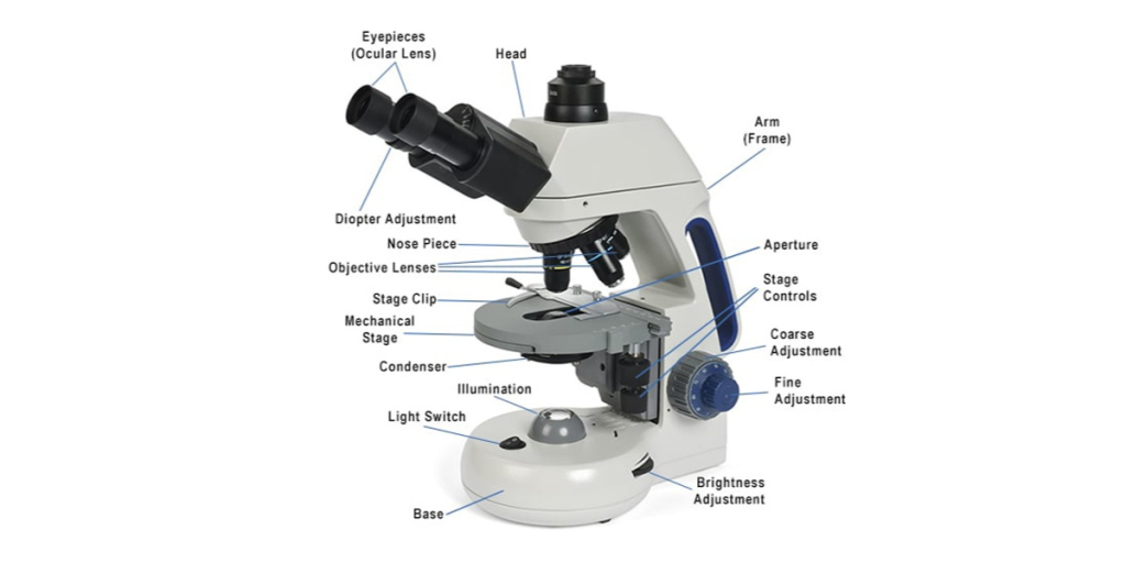
The microscope, a marvel of scientific ingenuity, has been instrumental in unraveling the mysteries of the microscopic world and reshaping our understanding of life itself. From its early origins as a simple magnifying glass to the sophisticated instruments of modern laboratories, the microscope has played a pivotal role in scientific discoveries, medical breakthroughs, and technological advancements. In this comprehensive exploration, we delve into the rich history, diverse types, applications, and the transformative impact of the microscope on various scientific disciplines.
The Early Quest for Microscope:
1. Antecedents:
The journey of the microscope begins with the ancient quest for magnification. In the 13th century, eyeglasses were crafted in Italy, and by the 16th century, the first magnifying lenses were combined to create early microscopes. These rudimentary devices laid the groundwork for the revolutionary strides to come.
2. Zacharias and Hans Janssen:
In the late 16th century, Dutch spectacle makers Zacharias and Hans Janssen are credited with creating the first compound microscope. This instrument featured a combination of lenses that significantly improved magnification, enabling the observation of smaller details.
The Rise of the Compound Microscope:

3. Galileo’s Contribution:
Galileo Galilei, renowned for his astronomical observations, applied his optics expertise to microscopes. Though his compound microscope had limitations, it marked an early effort to explore the microscopic realm.
4. Robert Hooke’s Micrographia:
In 1665, Robert Hooke published “Micrographia,” a groundbreaking work that showcased detailed illustrations of objects observed under a compound microscope. Hooke’s observations, including the iconic cork cells, opened a new chapter in the study of life at the microscopic level.
5. Antony van Leeuwenhoek:
A Dutch draper and scientist, Antony van Leeuwenhoek, achieved unparalleled success in microscope design. His single-lens microscopes, with remarkable precision, revealed a world of microorganisms previously unknown to humanity, fundamentally altering our perception of life.
Types of Microscopes:

6. Compound Microscopes:
Compound microscopes utilize multiple lenses to magnify specimens. They are widely used in various scientific disciplines, including biology, medicine, and materials science.
Brightfield Microscopes:
- Standard Illumination: Illumination from below the specimen produces a bright background, making stained or naturally pigmented samples visible.
Phase-Contrast Microscopes:
- Enhanced Contrast: Ideal for transparent specimens, phase-contrast microscopy enhances contrast without staining.
Fluorescence Microscopes:
- Fluorescent Labels: Used for visualizing structures that fluoresce when exposed to specific wavelengths of light, often employed in molecular and cellular biology.
7. Stereo Microscopes:
Stereo microscopes, also known as dissecting microscopes, provide three-dimensional views of larger specimens. They are commonly used for tasks such as dissection and inspection of intricate structures.
8. Confocal Microscopes:
Confocal microscopes use lasers to illuminate specific sections of a specimen, allowing for the production of detailed, high-resolution images. This technology is valuable in both biological and materials science research.
9. Electron Microscopes:
Electron microscopes utilize electron beams instead of light to achieve higher magnifications and resolutions. There are two main types:
Transmission Electron Microscopes (TEM):
- Internal Structures: TEMs transmit electrons through thin specimens, providing detailed views of internal structures at the nanoscale.
Scanning Electron Microscopes (SEM):
- Surface Imaging: SEMs scan electron beams across the surface of specimens, producing 3D images with exceptional detail.
10. Atomic Force Microscopes (AFM):
AFMs operate by scanning a sharp tip over a specimen, measuring the forces between the tip and the specimen’s surface. This technique is crucial for imaging at the atomic and molecular levels.
Applications Across Scientific Disciplines:
11. Biological Sciences:
Microscopes have been indispensable in advancing our understanding of cellular structures, microorganisms, and biological processes. They play a crucial role in fields like microbiology, histology, and genetics.
12. Medicine and Pathology:
In medicine, microscopes are essential tools for diagnosing diseases through the examination of tissue samples, blood smears, and other specimens. Pathologists rely on microscopy to identify abnormalities and make informed clinical decisions.
13. Materials Science:
Microscopy is fundamental in materials science for studying the structure, composition, and properties of materials at the microscopic and nanoscopic levels. This knowledge is crucial for developing new materials with specific functionalities.
14. Environmental Science:
Microscopes contribute to environmental science by aiding in the analysis of soil, water, and air samples. They enable researchers to study microorganisms, pollutants, and other components that impact ecosystems.
15. Physics and Nanotechnology:
In physics, microscopes are used to investigate the fundamental properties of matter and explore the behavior of particles at the atomic and subatomic levels. In nanotechnology, microscopes play a key role in manipulating and studying nanomaterials.
Technological Advancements:
16. Digital Microscopy:
The integration of digital technology has revolutionized microscopy. Digital microscopes feature cameras and imaging software, allowing for real-time observations, documentation, and sharing of images.
17. Artificial Intelligence in Microscopy:
Artificial intelligence is making significant inroads into microscopy, aiding in image analysis, pattern recognition, and even the identification of rare or abnormal structures in medical and biological samples.
18. Super-Resolution Microscopy:
Super-resolution microscopy techniques, such as stimulated emission depletion (STED) and structured illumination microscopy (SIM), surpass the diffraction limit, enabling the visualization of structures at the nanoscale.
FAQs: Navigating the Microscope
Q1: What is the diffraction limit in microscopy?
A1: The diffraction limit is a physical limit that restricts the resolution of traditional light microscopes. It is around half the wavelength of the illuminating light, limiting the smallest details that can be resolved.
Q2:
How does an electron microscope differ from an optical microscope?
A2: Electron microscopes use electron beams instead of light, allowing for much higher magnifications and resolutions. While optical microscopes are suitable for observing living cells and larger structures, electron microscopes excel at imaging smaller details at the nanoscale.
Q3: What is the importance of staining in microscopy?
A3: Staining is essential for enhancing the contrast of specimens, making specific structures more visible. Different stains target different cellular components, aiding in the identification of cells and tissues.
Q4: Can microscopes be used for educational purposes?
A4: Microscopes are commonly used in educational settings, from primary schools to universities. They facilitate hands-on learning and enable students to explore the microscopic world, fostering a deeper understanding of various scientific concepts.
Q5: How do you properly maintain a microscope?
A5: Proper maintenance includes keeping lenses clean, storing the microscope in a dust-free environment, and regularly checking for any signs of wear or damage. Following the manufacturer’s guidelines for specific microscope models is crucial.
Q6: Can microscopes be used to study living cells?
A6: Yes, certain types of microscopes, such as phase-contrast and fluorescence microscopes, allow for the observation of living cells. Specialized techniques, including time-lapse imaging, enable researchers to track dynamic processes within living cells.
Q7: Are there portable microscopes?
A7: Yes, portable microscopes are available, ranging from handheld devices to compact digital microscopes. These portable options are convenient for fieldwork, outdoor exploration, and situations where a traditional microscope might be impractical.
Q8: What is the significance of super-resolution microscopy?
A8: Super-resolution microscopy techniques surpass the diffraction limit of traditional microscopes, enabling researchers to visualize structures at the nanoscale. This is particularly crucial in studying cellular structures and nanomaterials.
Q9: How has digital technology impacted microscopy?
A9: Digital technology has transformed microscopy by allowing for real-time imaging, digital documentation, and sharing of microscopic images. It has also facilitated the integration of artificial intelligence for advanced image analysis.
Q10: Can microscopes be used for amateur microscopy?
A10: Absolutely. Entry-level microscopes designed for amateur microscopy enthusiasts are readily available. These microscopes are user-friendly, allowing hobbyists to explore the microscopic world in various fields, from biology to mineralogy.
Conclusion:
The microscope, a testament to human curiosity and scientific innovation, has unraveled the intricacies of the microcosm and transformed our understanding of the world. From the pioneering days of Zacharias Janssen to the cutting-edge technologies of today, microscopes have been indispensable tools across scientific disciplines. Whether revealing the hidden structures of cells, aiding in medical diagnoses, or advancing materials science and nanotechnology, the microscope continues to be a beacon of discovery. As technology progresses and new frontiers are explored, the microscope remains a symbol of humanity’s relentless pursuit of knowledge, offering a window into realms once invisible to the naked eye.
Related Links: The Thermometer


